
Research Toolkit

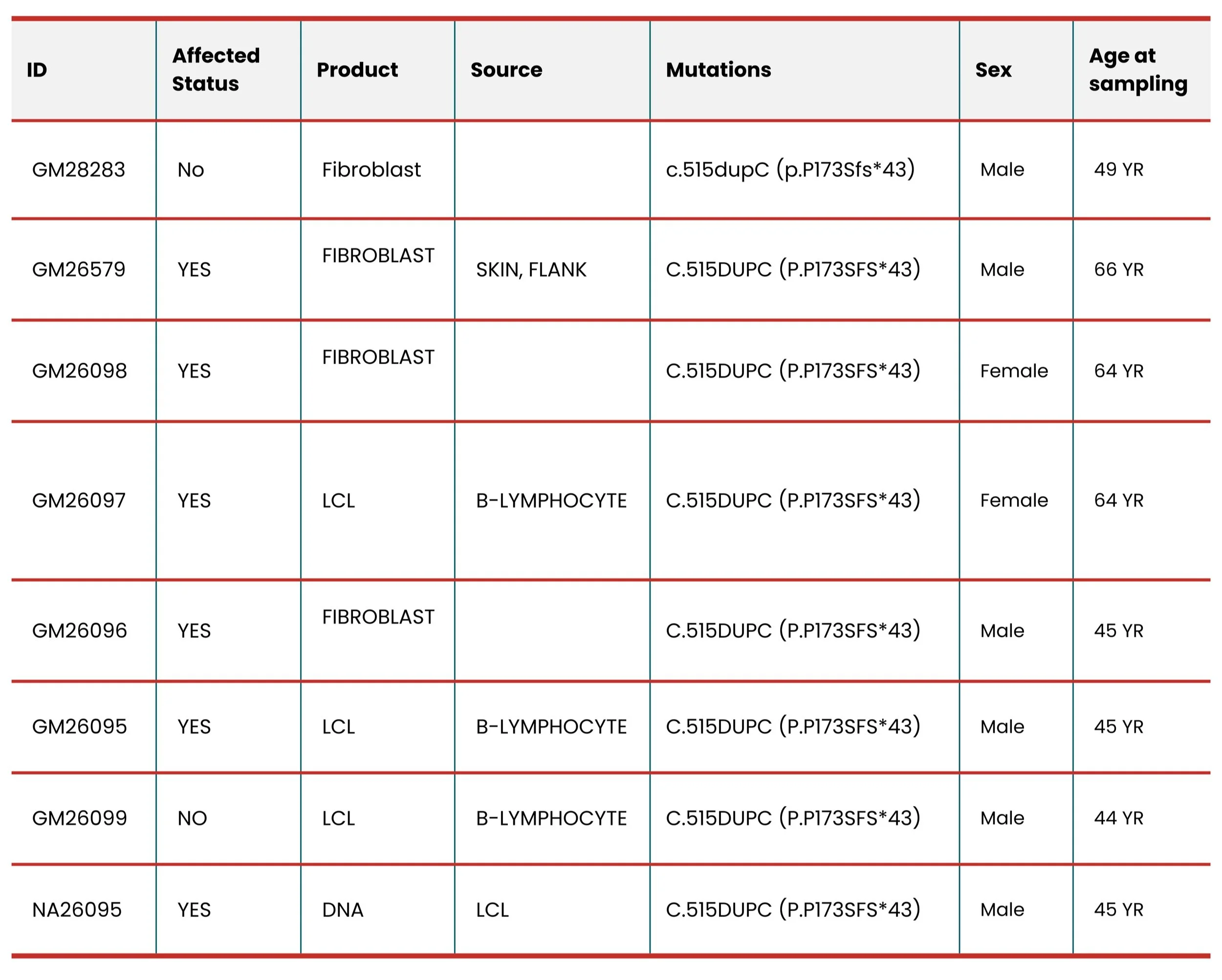
For researchers interested in studying MFM13, below is the list of resources currently available. While we are doing our best to make sure information in this table is up to date, please feel free to reach out to us for me most recent information.
Cells and DNA available from Coriell Cell Repository
Mouse Model
Partially humanized knock-in mouse model of MFM13
Generated at IIMCB, Warsaw, Poland (2025). Systematic phenotyping planned at the Czech Centre for Phenogenomics, Prague, Czechia.
This model is being developed to closely replicate the biological processes and symptoms seen in patients with MFM13, allowing researchers to explore how the HSPB8 mutation drives disease and to test potential therapeutic strategies.
Key characteristics:
- c.515dupC frameshift mutation in the HSPB8 gene, leading to an elongated C-terminal tail and toxic gain-of-function
- The C-terminal region is humanized - the mouse sequence is replaced with the equivalent human HSPB8sequence, ensuring the aberrant extension is identical to that found in patients
- Genetic background: C57BL/6JRj
- Phenotyping: not yet available (planned at the Czech Centre for Phenogenomics, Prague, Czechia)
Project Overview: Developing the First Humanized MFM13 Mouse Model
MFM13 in collaboration with IIMCB, Warsaw, Poland and Czech Centre for Phenogenomics, Prague, Czechia.
A validated animal model is a critical missing piece in MFM13 research. Without one, it is difficult to study disease mechanisms in living tissue, identify measurable endpoints, or test potential therapies. This page describes the scientific rationale, design, and current status of the first partially humanized MFM13 mouse model - a project led by Cure MFM13 in collaboration with International Institute of Molecular and Cell Biology (IIMCB, Warsaw, Poland) and the Czech Centre for Phenogenomics (CPP, Prague, Czechia). For background on the HSPB8 protein, disease pathology, and existing research tools, visit the HSPB8 Protein and Disease Pathology pages in our Research section.
1. Why a New Mouse Model Was Needed
MFM13 is caused by frameshift (fs) mutations in the last coding exon of the HSPB8 gene. All known mutations fall into one of two groups, defined by which alternative reading frame is used: one group produces an aberrant C-terminal extension of 19 amino acids (CE+19, known as fs2, Zhou et al., 2026), the other an extension of 49 amino acids (CE+49, known as fs3, Zhou et al., 2026). Both groups are thought to cause disease through the same core mechanism - the aberrant extension reduces protein solubility, drives aggregation, sequesters CASA complex members (BAG3, HSPA, STUB1), and impairs autophagic protein quality control (Zhou et al. 2026).
For this mouse model, c.515dupC (p.Pro173Serfs*43) was selected. It belongs to the CE+19 group and is one of the best-characterized pathogenic variant in MFM13 (Zhou et al. 2026; Tedesco et al. 2023 and 2025). Its CE+19 extension is the primary target for therapeutic strategies currently under investigation. An additional practical reason for this choice is that Cure MFM13 has developed an antibody specific to the c.515dupC (p.Pro173Serfs*43) mutant protein. This antibody recognises the aberrant CE+19 C-terminal extension and can distinguish the mutant HSPB8 from the wild-type protein. It will be a key tool during phenotypic characterisation of the mouse model, enabling direct detection and localisation of the mutant protein in muscle tissue.
The only previously available mouse model was a full knockout of Hspb8 (Jackson Lab, MMRRC_051194-JAX), which removes the protein entirely. A knockout cannot replicate the toxic gain-of-function mechanism, does not produce an aberrant C-terminal extension, and cannot be used to test therapies targeting the mutant protein or its downstream effects. A disease-relevant knock-in model was therefore urgently needed.
2. Protein Domain Structure and Solubility
2.1 Domain structure of HSPB8
Human HSPB8 is a 196 amino acid protein. It contains a conserved central alpha-crystallin domain (ACD) of 92 amino acids (G80 to V172), responsible for core chaperone activity and for binding the IPV motifs of co-chaperone BAG3, enabling CASA complex assembly. The ACD is flanked by a hydrophobic N-terminal region (NTR) containing the conserved RLFDQxFG motif, and a short polar C-terminal region (CTR, approximately 27 amino acids) whose hydrophilicity contributes a solubilising effect to the protein (Zhou et al. 2026). For a detailed description of HSPB8 protein structure and the CASA complex, see the HSPB8 Protein and Disease Pathology.
Mouse Hspb8 is highly conserved with the human protein: BLAST alignment of the WT proteins shows 94% identity over 196 residues, with the ACD and BAG3-binding function fully preserved across both species (Cure MFM13, unpublished analysis).

2.2 Solubility of WT versus mutant HSPB8: the case for humanization
Wild-type HSPB8 is a soluble protein whose short, polar CTR contributes to maintaining it in solution. The frameshift mutations replace this native CTR with an aberrant extension of reduced polarity and solubility. In silico analysis using the Protein-Sol tool shows that all HSPB8 frameshift (CE+19, fs2) mutant proteins have substantially reduced predicted solubility compared to wild-type: the WT whole-protein score is 0.678, while c.515dupC scores 0.345 - a reduction of approximately 49% (Zhou et al. 2026). The native CTR (fs1) has an acidic isoelectric point (pI 1.00) and net charge of -4.1 at pH 7, whereas most CE+19 (fs2) mutant CTRs shift to a basic isoelectric point (pI ~10.95) and net charge of +2. AlphaFold structural modelling further predicts an additional alpha-helix in the CTR of all mutant proteins, absent in WT, likely contributing to aberrant interactions and aggregation (Zhou et al. 2026).
Experimentally, Tedesco et al. (2023) confirmed that human HSPB8 frameshift (fs2 and fs3) mutants are highly insoluble: NP-40 fractionation showed mutant proteins partitioning preferentially into the insoluble fraction, and molecular dynamics simulations revealed an approximately 3-fold increase in aggregation propensity versus WT. This aggregation seems to bean intrinsic property of the mutated amino acid sequence, independent of CASA member interactions (Tedesco et al. 2023).
2.3 Why introducing the frameshift into the mouse sequence is not sufficient
A critical design question was whether simply introducing c.515dupC into the mouse Hspb8 sequence would reproduce the human disease mechanism. It would not: the aberrant C-terminal extension is determined by the downstream sequence context at the frameshift site, and because the mouse and human sequences differ in this region, the same frameshift produces a different amino acid extension in the mouse than in human patients.
To assess the consequence of this difference, solubility was predicted for all four protein variants using two independent in silico tools: NovoPRO and Protein-Sol (Cure MFM13, unpublished analysis). These tools approach solubility from different angles. NovoPRO analyses specifically the last 20 amino acids of the protein - the region most directly altered by the frameshift - making it particularly sensitive to changes in the C-terminal extension. Protein-Sol assesses the whole protein sequence, with scores above 0.5 indicating predicted soluble expression (Figure 1b).
The results are consistent across both tools and tell a clear story:
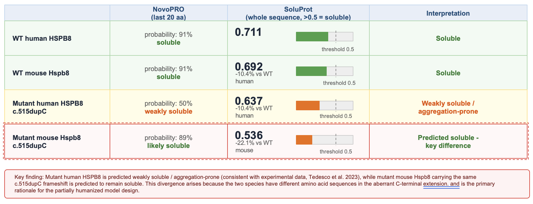
Both wild-type proteins are confidently predicted soluble by both tools. The mutant human protein shows a marked deterioration: Protein-Sol drops from 0.678 to 0.345, falling well below the 0.5 solubility threshold - a reduction of approximately 49%. This is consistent with the experimental insolubility of human mutant HSPB8 demonstrated by Tedesco et al. (2023), who showed by NP-40 fractionation and filter retardation assay that human frameshift mutants partition preferentially into the insoluble fraction and form high molecular weight aggregates.
The NovoPRO result is particularly informative: because it focuses exclusively on the last 20 amino acids, it directly captures the chemical character of the aberrant C-terminal extension - the part of the sequence that differs between human and mouse. The human mutant drops from 91% to 50% probability of solubility, directly reflecting the hydrophobic character of the CE+19 extension. The mouse mutant, in contrast, remains at 89% - indicating that despite carrying the frameshift at the same genomic position, its C-terminal extension has a fundamentally different chemical character (1b-c).
The Protein-Sol score for the mouse mutant (0.479) does fall slightly below the 0.5 threshold, suggesting some reduction in overall solubility. However, combined with the NovoPRO result of 89%, the overall prediction is that the mouse mutant protein would remain largely soluble. This is in stark contrast to the human mutant, where both tools consistently predict reduced solubility and aggregationpropensity. If the mouse mutant does not aggregate, the core pathogenic mechanism - sequestration of CASA complex members and proteostasis failure - would likely not be reproduced, and the model would display little or no disease phenotype despite carrying the frameshift at the same genomic position.
This is the primary justification for the partially humanized design: by replacing the mouse sequence with the human HSPB8 sequence in this region, the model ensures the c.515dupC frameshift produces the same insoluble, aggregation-prone CE+19 extension as in patients.

3. The Partially Humanized Mouse Model
3.1 Design rationale
Rather than introducing a point mutation into the mouse gene, the 3’ end of the mouse Hspb8 coding sequence was replaced with the corresponding human HSPB8 sequence carrying c.515dupC. This ensures: (i) the aberrant CE+19 extension is encoded by the same human sequence as in patients, with the same amino acid composition and predicted insolubility; and (ii) the model is directly suitable for testing human-sequence-targeting therapies such as antisense oligonucleotides (ASOs), which would not bind the equivalent mouse sequence.
3.2 CRISPR/Cas9 strategy
A gRNA (ATTTGCCTCGCTTTCTCCAG, PAM: AGG) targeting exon 3 of mouse Hspb8 was used to introduce a double-strand break. A dsDNA donor was provided for homology-directed recombination, comprising:
- 60-nucleotide left homology arm
- Human hHSPB8 coding sequence (167 nt of the 3’ end, including c.515dupC and the CE+19 extension)
- Two silent mutations (A>T at position 62, G>A at position 65 of the donor) to disrupt the PAM sequence and prevent repeated Cas9 re-cutting
- SV40 poly(A) signal-containing sequence to facilitate transcription termination
- 60-nucleotide right homology arm
The resulting transcript encodes a chimeric mRNA: the upstream portion is of mouse origin, while from approximately codon 117 onward the sequence is human, carrying c.515dupC and the CE+19 extension. The model was generated on a C57BL/6JRj genetic background by dr Michał Brouze at IIMCB, Warsaw, Poland (mutagenesis strategy is decribed here 2025.GEU.68).
Phenotyping will be conducted on heterozygous animals - carrying one mutant Hspb8 allele and one wild-type allele - selected through breeding following initial line generation. This mirrors the genetic situation in MFM13 patients, where the disease is inherited in an autosomal dominant manner and all identified cases are heterozygous carriers. The presence of the wild-type allele is also mechanistically relevant: the mutant HSPB8 protein retains the ability to homodimerize with wild-type HSPB8 and sequester it into aggregates, exerting a dominant negative effect on the functional CASA complex (Zhou et al. 2026; Tedesco et al. 2023).
3.3 CASA complex binding in the model
The ACD - which mediates substrate recognition and BAG3 binding - is retained from the mouse sequence upstream of the CRISPR cut site. The human sequence begins approximately at aa 117, within the latter part of the ACD and into the CTR region. The frameshift and CE+19 extension are fully human and identical to the patient's protein. Researchers should account for this chimeric nature when designing and interpreting CASA interaction studies in this model.
3.4 Future ASO therapy readiness
Because the inserted human sequence extends upstream of c.515dupC, the model includes a stretch of wild-type human coding sequence before the frameshift. ASO therapies designed against the human HSPB8 sequence could potentially be tested directly in this model - which would not be possible in a standard mouse model where the target sequence differs. This should be confirmed with the scientific team before designing ASO studies.
4. International Collaboration
The MFM13 mouse model was developed through a three-way international collaboration between Cure MFM13, the International Institute of Molecular and Cell Biology (IIMCB) in Warsaw, Poland, and the Czech Centre for Phenogenomics (CCP) in Prague, Czechia.
4.1 IIMCB, Warsaw, Poland
IIMCB was established in 1997 under an international agreement between the Government of Poland and UNESCO, giving it a unique legal status in the Polish scientific system. It holds the highest scientific category (A+) from the Polish Ministry responsible for science and is a member of the EU-LIFE alliance of independent European research institutes. IIMCB is recognized for its work in structural biology, RNA biology, cell biology, and neurodegeneration. Mouse model generation was carried out by Dr Michał Brouze Team at IIMCB.
4.2 Czech Centre for Phenogenomics (CCP), Prague, Czechia
CCP is part of the Institute of Molecular Genetics of the Czech Academy of Sciences and is one of Europe’s largest phenogenomicsinfrastructures, employing approximately 140-145 specialists and handling more than 1,400 user requests annually from over 300 academic and industrial partners. CCP is a member of INFRAFRONTIER and a contributor to the International Mouse Phenotyping Consortium (IMPC). Phenotyping of the MFM13 model at CCP is conducted through the RD-Factory Program, within which MFM13 was selected in the 3rd Call for Nominations (2026)

5. Phenotyping Goals at CCP
Comprehensive phenotyping of the MFM13 model at CCP aims to establish the model as a validated tool for translational research and future therapeutic development.

5.1 Goals in detail
1. Confirm disease-relevant phenotypes. Assess muscle pathology including weakness, rimmed vacuoles, myofibrillar disorganisation, HSPB8-positive aggregates, and CASA pathway markers (SQSTM1/p62, TDP-43, ubiquitin).
2. Map disease onset and progression. Age-stratified studies to determine when phenotypes first appear and how they progress, establishing a disease timeline for therapeutic window design.
3. Identify translational readouts. Functional (grip strength, rotarod, electrophysiology), molecular (protein aggregation markers, autophagy flux), and behavioural endpoints suitable as outcome measures in future drug trials.
4. Generate preclinical data. Robust, standardised datasets to support future therapeutic testing and regulatory submissions where applicable.
5. Enable ASO and gene therapy testing. The humanized sequence design enables future testing of human-targeting antisense oligonucleotide and gene therapy approaches directly in this model.
6. Open community access. The mouse model and phenotyping data will be made available to the broader research community to accelerate global MFM13 research.
5.2 Future Phenotypic
First, we are planning to perform a general phenotypic characterization to evaluate the differences between wild-type (WT) and MFM13 mouse models. Based on the results, we will expand the study to include more detailed phenotypic analyses.

6. Current Status and Contact
As of June 2026, the mouse line is currently being generated at IIMCB in Warsaw, Poland. Systematic phenotypic characterization at CCP in Prague, Czechia, is planned to follow. Updates will be posted at curemfm13.org and on social media as the project progresses.
Researchers interested in collaboration or future access to the model are invited to contact:
Dr Anna Kordala, Program Director, Cure MFM13 and dr Sylwia Szwec, Research Program Manager
ania@curemfm13.org | sylwia@curemfm13.org | curemfm13.org
References
- Zhou W, Marchesi V, McLeod M, Kordala AJ, Szwec S, Mielcarz JA, Poletti A, Tedesco B (2026). Molecular, cellular, and clinical aspects of myofibrillar myopathy caused by HSPB8 frameshift mutations. BBA - Molecular Basis of Disease 1872:168244. doi:10.1016/j.bbadis.2026.168244. [Primary source for this document.]
- Tedesco B et al. (2023). HSPB8 frameshift mutant aggregates weaken chaperone-assisted selective autophagy in neuromyopathies. Autophagy 19(8):2217-2239. doi:10.1080/15548627.2023.2179780.
- Tedesco B et al. (2025). Novel HSPB8 mutations in severe early-onset myopathy with involvement of respiratory and cardiac muscles cause proteostasis defects in cell models. Eur J Hum Genet 33:1015-1024. doi:10.1038/s41431-025-01868-z.
- Mutagenesis strategy 2025.GEU.68, prepared by Michał Brouze, IIMCB, Warsaw, Poland (July 2025).
- In silico solubility analysis (NovoPRO), Cure MFM13, unpublished analysis. BLAST sequence alignments (WT and mutant human vs mouse HSPB8): Cure MFM13, unpublished analysis.
